Advanced technologies in audiology and ENT diagnostics
We are UAE based company specializing in the distribution of hearing aids and medical equipment to clinics, hospitals and audiology centers, with the provision of fast local technical support and reliable after-sales services.
Advanced technologies in audiology and ENT diagnostics
We are UAE based company specializing in the distribution of hearing aids and medical equipment to clinics, hospitals and audiology centers, with the provision of fast local technical support and reliable after-sales services.
We are a company specializing in the distribution of medical devices and equipment in the United Arab Emirates, with a primary focus on audiology technologies, hearing aids, and ENT diagnostic devices. We partner with leading global companies to provide reliable medical devices and technologies that help doctors and audiology specialists deliver the best possible patient care. Our goal is to offer integrated solutions that combine advanced medical technology with professional technical support to ensure optimal outcomes for patients and healthcare providers. Our services include:
Distribution of Advanced Hearing Aids
Provision of Audiology Diagnostic Devices
Supporting Clinics and Hospitals with the Latest Medical Technologies
Training, Technical Support, and After-Sales Service
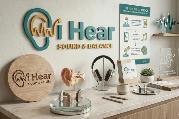
Thehearing aids we offer:
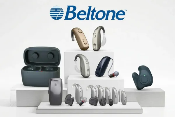
At iHear, we provide advanced hearing aids powered by cutting-edge technology and intelligent AI-driven sound processing, delivering exceptional clarity, comfort, and personalized performance. Our solutions are powered by Beltone, a global leader in innovation. Discover the latest Beltone range, including Envision, Boost Max, and Commence—designed to automatically adapt to your environment, enhance speech understanding, and provide a natural listening experience in every situation. From discreet RIC to powerful BTEdevices, our hearing aids are tailored to meet all levels of hearing needs, ensuring effortless control, seamless connectivity, and a better quality of life through improved hearing.
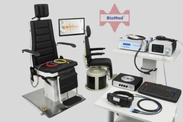
We provide comprehensive solutions for hearing and balance diagnostics, combining German precision with technological innovation. The catalog includes advanced audiological systems such as eAUDIOUSB and eTYMPUSB for hearing and middle ear assessment, in addition to eABRUSB and eOAE technologies for advanced testing and newborn screening. In the field of balance, systems like eVNGUSB and eHITUSB stand out for precise analysis of eye movements and semicircular canals, alongside KALORIstar thermal irrigation devices and the ePOSTURO system for posture analysis. All devices are integrated with the unified Diagnostic Manager(eDM) software to efficiently manage patient data, ensuring a professional workflow and accurate clinical diagnosis.
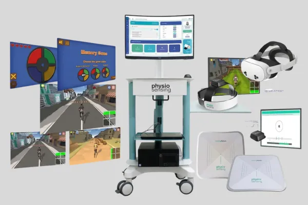
The PhysioSensing OtoneuroSystem and PhysioSensingPosturography Station are advanced solutions in the field of balance disorder assessment and treatment. The OtoneuroSystem provides a comprehensive platform that combines balance analysis, motion sensors, and virtual reality for accurate diagnosis and interactive therapy. Meanwhile, the Posturography Station focuses on high-precision balance measurement through a Force Plate platform, analyzing stability indicators and fall risk. Both systems support clinicians in making precise decisions, improving treatment outcomes, and delivering a better patient experience.